Message Boards Message Boards

How is the sample 3D image of Knee is created?

The sample 3D image of knee in Mathematica is the MRI of Markus van Almsick(https://www.youtube.com/watch?v=s1ot5MltxM8&spfreload=1).

The corresponding image dimension is {128, 128, 128} and I can clearly have all the three views (sagittal,axial and coronal) from the same image.

However, normally MRI consists of 20-25 2D slices and I have separate image slices. But I want to have my 3D image containing all the three views (sagittal, axial and coronal) as there are for the sample knee image in which it seems that all the slice sets along the orthogonal directions are somehow combined to create this view.

Therefore, I would like to know how this image was re-sized with the above mentioned dimensions?

POSTED BY: Subhrajyoti Maji
2 Replies

I will do this for the engine example.

If you have slices:

dataimg = Import["ExampleData/CTengine.tiff"]

enter image description here

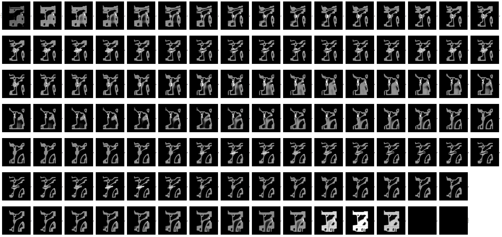
You combine them using Image3D (it does not really depend on the image dimensions of the individual images.

Image3D[dataimg]

enter image description here

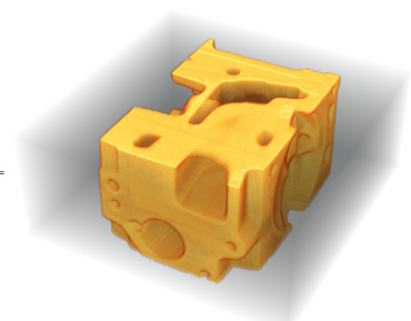
You can change the box ratios by:

Image3D[dataimg, BoxRatios -> {1, 1, 2}]
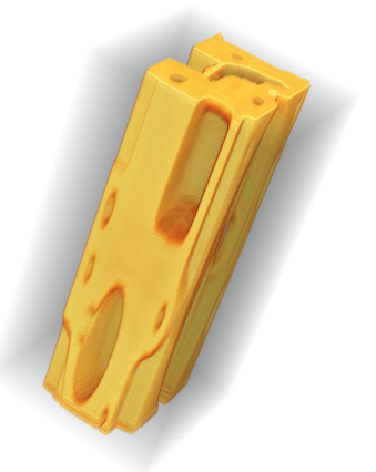
enter image description here

You can also use

ImageResize[Image3D[dataimg], {300, 300, 150}]

to resize the image.

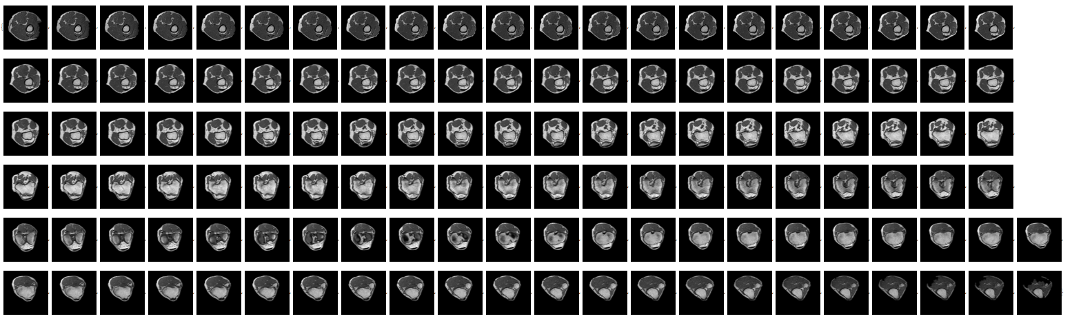
You will see that this is the same principle for the knee. If you use

Image3DSlices[ExampleData[{"TestImage3D", "MRknee"}]]

you will see the individual slices of the knee. Image3D reconstructs a 3D image from that.

enter image description here

Cheers,

Marco

POSTED BY: Marco Thiel

@Marco Thiel

Thanks Marco for your kind information. Now I got it.

POSTED BY: Subhrajyoti Maji
Reply to this discussion
Community posts can be styled and formatted using the Markdown syntax.
Reply Preview
Attachments
Remove
or Discard

Group Abstract Group Abstract